
Environmental links aside, there are several cell types and proteins that malfunction during the onset of SLE. One main protein that is malfunctioning is the production of autoantibodies. Autoantibodies are antibodies that mistakenly target and react with a person’s own tissues or organs. The most common form, antinuclear antibodies (ANA), is found is almost 95% of all SLE patients. Autoantibodies are produced by B cells through random production or in a response to a foreign protein or substance entered into the body. Either way of production leads to this production of pathological autoantibodies, that cause cell apoptosis and lysing depending on the autoantibody. What makes autoantibodies so damaging to the host is that they attack and target nucleic acid containing particles; meaning the hosts cells which hold DNA and other cells are directly targeted by autoantibodies and are destroyed. An example of this destruction of DNA is the specific autoantibody Anti-dsDNA which target double stranded DNA in the host. It is not truly known why these nuclear components become autoantigens; however, it is thought to be connected with cell apoptosis. Cell apoptosis is strictly regulated and contains a series of pathways leading to the regulated removal of unwanted cells such as these described autoantibodies. In the event this pathway becomes impaired, it could lead to the release of modified nuclear structures that would trigger this downstream immune response leading to the production of autoantibodies. This also will lead to the destruction of chromatin which is an autoantigen that is clustered in apoptotic cell clusters. This ultimately releases increased levels of soluble Fas receptor (apoptosis antigen 1), bcl-2 and polymorphisms in cell death and transcription factors.
Two other autoantibodies that is important to mention that are malfunctioning is the anti-histone antibodies and anti-SSA/Ro autoantibodies. Anti-histone antibodies specifically target histone protein subunits or histone complexes including histone protein subunits, H1, H2A, H2B, H3 and H4. This is incredibly damaging because histones are critical in packing DNA into the cell and into chromatin and chromosomes and the regulation of gene production and gene expression. Anti-histone antibodies that attack these histones can cause mutated gene expression which could result in damaged DNA cells and create another downstream effect that leads to chronic inflammation and permanent damage of connective tissues in a lupus patient. It is thought that antihistone autoantibodies are triggered through viral infections and certain drugs. Anti-SSA/Ro autoantibodies function similarly but they attack Ro proteins (Ro52 and Ro60). Anti-SSA/Ro activity occurs on the cell surface and bind to Ro proteins expressed on the cell membrane. Anti-Ro/SSA is produced in the cytoplasm of cells in the epidermal layer of the skin following some form of UV radiation or dendritic cell damage. Simultaneously, Ro antigens are upregulated on the cell surface resulting in marking cells for apoptosis. It is important to note that alleles in MHC II (HLA-DR3 and HLA-DR2) are associated with T-cell response and play a critical role in Anti-Ro/SSA antibody formation. This is important because MHC II is a set of closely linked polymorphic genes that are essential for the adaptive immune system, meaning that now both the adaptive and innate immune system are affected and changed during the disease process of SLE. In patients with SLE, high levels of Anti-Ro/SSA are correlated with elevated levels of IFN-alpha, which is mainly involved in an innate immunity against viral infection.
In patients with SLE, TLR7 and TLR9 are genetically impacted and mutated to further cause inflammation and degradation to human cells. TLR7 has a receptor TREML4 that is an essential positive regulator of TLR7 signaling. Research has shown that inn TREML4 -/-, mice macrophages that are hyporesponsive to TLR7 agonists, macrophages fail to produce type I interferons due to impaired phosphorylation of the transcription factor STAT1 by the mitogen-activated protein kinase p38 and decreased recruitment of the adaptor Myd88 to TLR7. TREML4 deficiency reduced the production of inflammatory cytokines and autoantibodies in MRL/lpr mice, suggesting that TLR7 is a vital component of antiviral immunity and a predecessor factor in the pathogenesis of rheumatic diseases such as SLE. In TLR9, viruses like EBV can genetically alter the expression of TL9. This can cause transcription and translation issues that results in lack of pro-inflammatory regulations. This loss of TLR9 exacerbates progression of SLE and leads to increased activation of dendritic cells. TLR9 also controls the release of IgA and IFN-a in SLE, and loss of the receptor leads to higher levels of both molecules. In SLE, TLR9 and TLR7 have opposing effects. TLR9 regulates inflammatory response, while TLR7 promotes inflammatory response.
One cell/pathway that is mutated and altered through this disease is the activation of memory B cells in the absence of cognate T-cell help. Research through Doreau et al, shows that inflammation can perpetuate autoimmunity through cytokine driven activation of memory B cells. In patients with SLE, a pathological response to nuclear antigens creates somatic mutations leading to increased affinity of the autoantibodies through switching to IgG. This IgG switch contains IgG antinuclear autoantibodies that activate TLRs within plasmid dendritic cells (DCs), monocytes and B cells. This TLR engagement induces transcription of proinflammatory mediators including type I interferons which activate myeloid DCs to coactivate T cells. This release both BAFF and IL-6 which are required for the induction of Th17 cells and help stimulate DC maturation, TLR expression and IL-6 release (BAFF). This TLR expression in B cells increases BCR-mediated signaling and results in the survival of B cells that internalize nucleic acid-containing immune complexes. This B cell pathway ties in a lot of genetic mutations that can occur in patients that test positive for SLE and explains the critical role that pathogenic IgG autoantibodies play in tissue inflammation and these multiple different types of pathways in SLE.
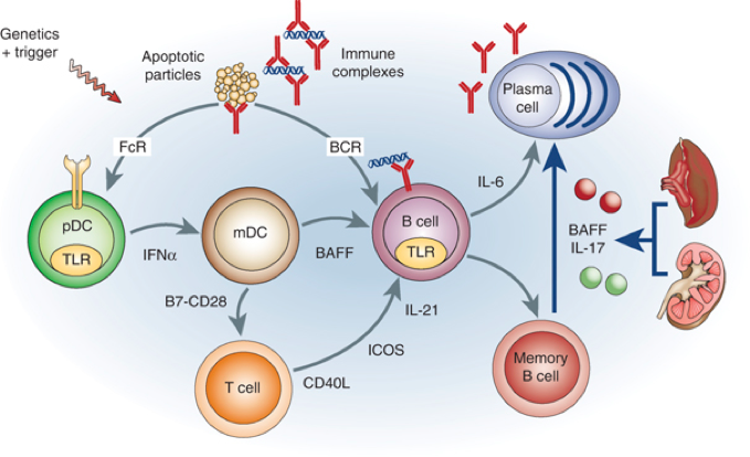
Seen in the picture above recognizes a new pathway for memory B-cell activation in systemic lupus erythematosus (SLE). Multiple amplification pathways (shown in gray) are activated in SLE (Doreau et al). The novel NF-B-dependent pathway (shown in blue) circulates cytokines released from inflamed organs induce differentiation of memory B cells to plasma cells thus perpetuating the autoimmune response. This picture is just emphasizing how complex SLE is and how all of these genetic mutations are linked/how these autoantibodies all play a role in this autoimmune disorder. On top of this picture and research emphasizing the interconnected roles of memory B cells, macrophages and dendritic cells are being impacted and altered because of this disease. It is also emphasizing the contribution of IL-17 and how chronic inflammation can occur. In human SLE, IL-17 is released by both CD4+ and CD3+ T cells and by effector T cells in inflamed organs (like the kidney or liver) to enhance other proinflammatory mediators. While IL-17 research is still underway, the production of both CD4+ and CD3+ also creates a downregulated response in the production of macrophages and dendritic cells.
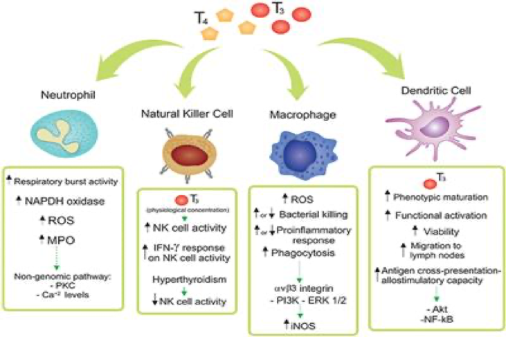
The picture above shows a simple diagram on how the production of CD4+ and CD3+ help T cells create macrophages and dendritic cells and also their function once activated. Increased production from CD4+ and CD3+ lead to the overproduction of these cells, which ultimately leads to an overactivation of the immune system. This creates an overabundance in proinflammatory responses and results in chronic inflammation.
Finally, it is important to briefly explain the aggregation of IgG antibodies and this type III hypersensitivity reactions seen in SLE. In patients with SLE, there is an overaccumulation of IgG antibodies, which are insoluble immune complexes that form in the blood and are deposited in various connective tissues.
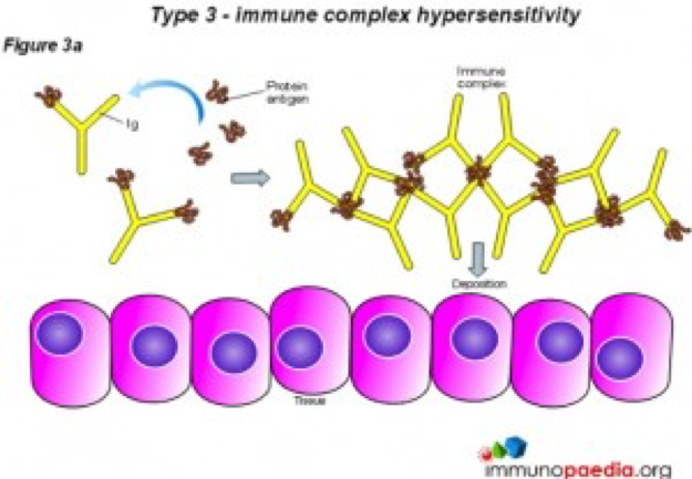
As seen in the picture above, IgG is deposited onto connective tissues which could ultimately lead to an immune response following the classical pathway of the complement system. As stated, earlier genes that regulate the complement system are found as defective in SLE patients and can lead to further tissue damage. The IgG antibodies bind to the tissue and start this accumulation onto the tissue surface. This large accumulation cannot be removed or cleared by the host normally so inflammatory mediators and other cells like neutrophils, basophils and eosinophils are recruited the affected tissue. This leads to chronic inflammation, tissue damage and the influx of phagocytes that engulf and destroy potentially healthy tissue.
So, the larger question is since there is a large and complex immune response in SLE occurring in a patient, why is the immune system behaving this way? The main cause of this is in fact genetic factors present in SLE patients. Along with rare high-risk mutations seen in the complement pathway, mutations in genes encoding nucleases in response to type I IFNs and genetic variants of IRF5 and IRF7 which are involved in signaling thorough endosomal (DC) TLRs activated by DNA or RNA are other examples of variants that can be mapped to molecular pathways responsible for innate immune activation. With IRF5 there is a common haplotype that is expressed in pDCs and B cells that is associated with an increased risk of SLE. Seen in Figure 1, it clearly shows this interaction of pDCs and B cells but does not highlight the genetic IRF5 mutation. IRF5 also acts downstream of the TLR-MyD88 signaling pathway in the induction of proinflammatory cytokines. A final gene variant seen in SLE patients contributes to altered thresholds for lymphocyte activation or efficiency of immune cell signaling. This haplotype is important in determine whether anti-DNA autoantibodies are produced. Overall while there are many complex systems that are mutated and altered in SLE, the general trend observed is that through a series of joint mutations leads to this cascade effect causing overall hypersensitivity and chronic inflammation in joints and connective tissues. Needless to say SLE is not only multifaceted but also deeply complex in its formation and overall function.
